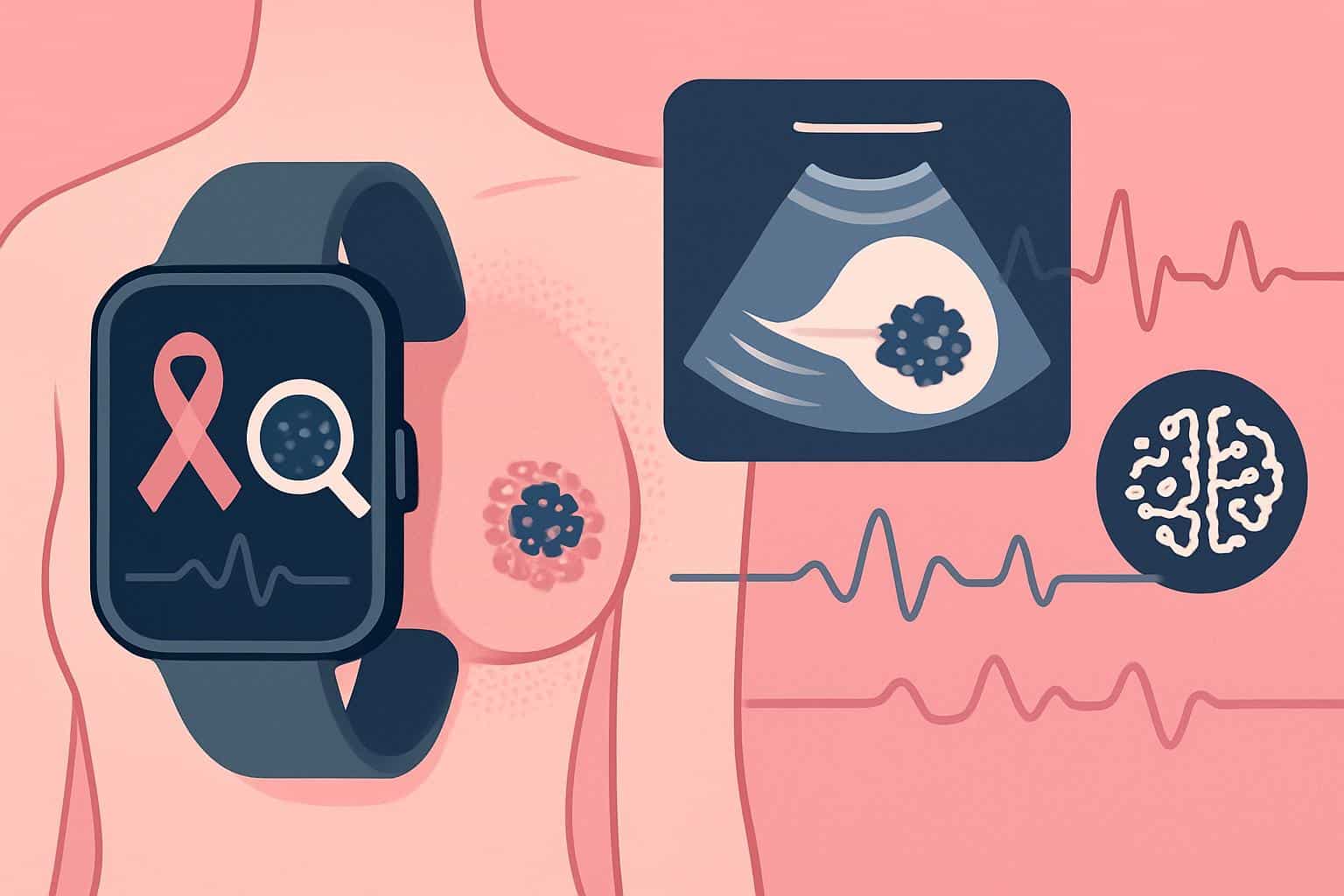
Step counters and sleep trackers are entering hospitals, promising resolution of a very high-stakes question: In which patients does surgery provide a cure? Using continuous sensing combined with artificial intelligence to flag subtle changes long before symptoms appear, researchers are developing wearable health monitors that could serve as “picks and shovels” in the future “gold rush” for rapid and predictive risk assessments for early breast tumors between clinic appointments.
Among the most watched: that of MIT associate professor Canan Dagdeviren, whose lab has engineered a flexible, ultrasound-based patch designed to image breast tissue while worn. The idea is simple yet brilliant: translate biological signals into electrical code that can be analyzed on the fly, moving imaging out of the radiology suite and into everyday life.

Why Continuous Breast Screening Matters for Outcomes
Breast cancer is the most frequent cancer in women worldwide. The American Cancer Society estimates that about one in eight women will be diagnosed with it over the course of her lifetime, and more than 40,000 women die from the illness annually in the United States. Survival depends on catching tumors early, when they are small and localized.
Mammography is the best standard of care and saves lives, but it’s not perfect. Sensitivity is reduced in women with dense breast tissue, and intervals also allow tumors to develop between scheduled screenings. These so-called “interval cancers” are often more aggressive and associated with worse outcomes, according to studies cited by journals including JAMA and the BMJ.
“How am I doing?” Rather than waiting a year or two between images, a wearable device could detect a growing abnormality within days or weeks, leading to the diagnostic workup happening sooner. This could be particularly useful for high-risk women or women with dense breasts — who now receive density notifications under updated FDA rules.
From Smart Bras to Wearable Ultrasound Patches
The MIT patch is part of this broader wave of hardware innovation. The bottom line: The device shapes itself to each woman’s breast, glides in a “track” within a cushioned singlet, and uses ultrasound rather than radiation. Initial lab and pilot human data, reported in the journal Science Advances, support the feasibility of detecting small lesions with reduced operator dependence.
Different teams are looking into different signals. Thermal methods examine minuscule temperature asymmetries related to tumor-induced blood vessel formation; firms like Cyrcadia Health have trialed smart patches that track circadian thermal profiles. Niramai’s AI-aided thermal imaging in India has targeted improving the availability of cost-effective screening tests in community settings. Handheld devices such as iBreastExam deploy soft piezoelectric sensors to map tissue stiffness, providing an indicator of abnormalities with no radiation involved, and are suitable for point-of-care screening.
None of these tools are meant to replace traditional mammography or MRI for diagnosis. They might instead serve as an early warning layer, elevating people to imaging and perhaps biopsy when the data indicate cause for concern, and providing reassurance when signals remain stable.
AI Translates Signals Into Early Warnings
Data is the unlock. Wearables produce continuous streams — ultrasound frames, temperature trends, impedance maps, motion-adjusted baselines — that machine-learning models can read at scale. By getting to know a person’s “normal,” algorithms can pick up deviations that might signal a developing mass, inflammation, or response to treatment.

Modern AI also helps drive down two important risks: false positives that create anxiety and unnecessary procedures, and false negatives that miss disease. Heterogeneous ensemble-trained models validated on Indian gold-standard imaging can be adapted to trigger high-specificity alarms while not losing sensitivity. Methods like federated learning can boost accuracy across health systems without consolidating sensitive data.
AI-powered triage, importantly, might also personalize surveillance. Among other tools, the U.S. Preventive Services Task Force recommendations establish a standard baseline for mammography, but wearables and algorithms can potentially refine how often and when adjunct screening should be pursued by those at higher risk, such as patients with genetic predilections or prior abnormal results.
What Needs to Be Shown Before Widespread Use
Clinical validation is paramount. The devices will have to demonstrate that they are able to detect clinically meaningful lesions earlier than the current practice does, and that such earlier detection would lead to better outcomes — not just more biopsies. Prospective randomized or well-controlled studies including multiracial populations and dense-breast cohorts are imperative.
Regulators will look for similar attention to safety and performance across settings, and credible cybersecurity and privacy protections. Reimbursement is another barrier; payers generally need proof that the new screening pathways bring better health results and are cost-effective relative to typical care.
Equity needs to be incorporated from the beginning. When the distribution of training data is not balanced across different groups, the predictions made are less likely to be good for those underrepresented groups. On the flip side, well-designed wearables might increase access by putting high-quality screening right in primary care clinics, pharmacies, or rural and resource-poor settings.
What This Means for Patients and Their Care Teams
Picture an at-risk woman tapping on a comfy patch for a few minutes once a week. If the device’s AI detects an abnormal pattern, her care team gets a notification and sets up imaging for a diagnosis immediately — weeks or months before her next routine mammogram. For patients on treatment, wearables could monitor tumor response in near real time, indicating whether to persist or change treatment.
The result is a more intelligent and continuous safety net around breast health. As MIT and others move forward into human studies, the mission is clear: Enhance today’s screening with unobtrusive technology that catches cancer at its earliest curable juncture — and only escalates when the data says it should.

